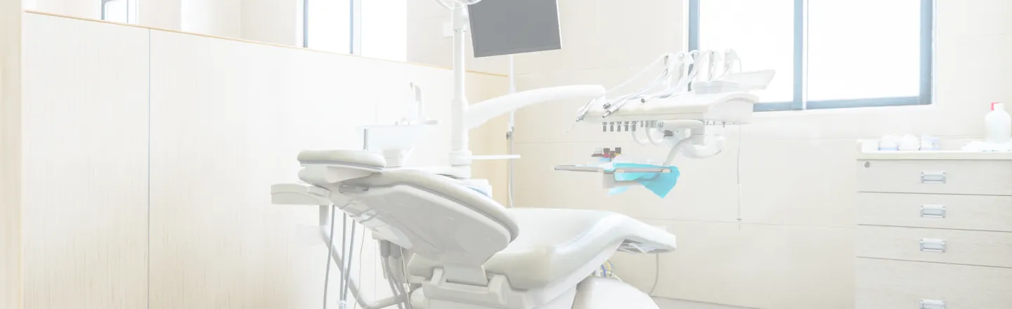
Intraoral camera in [city], [st] for clearer exams
At [practice_name] in [city], [st], an intraoral camera helps make dental exams clearer and easier to understand. The intraoral camera in [city], [st] is a small handheld device that captures magnified, high-resolution images of your teeth and gums. These images display on a chairside screen so concerns are easier to see, discuss, and monitor over time.
Intraoral camera explained
An intraoral camera is a pen-sized wand with a tiny lens and LED lights. It records live video and still photos inside the mouth, producing digital dental images that show details that can be hard to see with mirrors alone. Clear visuals support precise documentation and make conversations about findings more straightforward.
Patients often ask, “What is an intraoral camera and why is it used?” The tool helps reveal plaque buildup, early cavities, cracks, worn fillings, gum inflammation, and other changes in hard-to-reach areas. It is not a replacement for X-rays. Intraoral camera vs X-rays is a common comparison, and both are complementary. X-rays show what lies between and inside teeth and bone, while the camera shows color, texture, and surface details.
Benefits of using an intraoral camera
- Seeing what [dr_type] sees improves chairside patient education and builds confidence in care decisions.
- High magnification may help with early cavity detection and documentation of small cracks or chips.
- Digital photos create a visual record to track changes over time and evaluate how treatments are working.
- Images can be shared securely with specialists if a referral is needed for coordinated care.
- No radiation is involved, and the process is comfortable for most patients.
- Clear visuals can support discussions about home care strategies tailored to your mouth.
How an intraoral camera works
Patients often search “How does an intraoral camera work?” The device uses bright, cool LED lights and a tiny sensor to capture images while the dental team gently moves the wand around your mouth. A disposable barrier cover is used for each patient to protect your health. The live image appears instantly on a monitor, and still photos are saved directly to your secure dental record.
During a routine exam at our [city], California practice, the camera may be used to document stained grooves, fractured edges, leaky fillings, or inflamed gum pockets. Clear images make it easier to compare areas at future visits and to discuss options if treatment is recommended.
What to expect at your visit
The intraoral camera exam is quick and noninvasive. You will sit back as the wand gently scans your teeth and gums. Most patients feel only light pressure. No special preparation is required, and there are no restrictions after the appointment. If a concerning area is shown on the screen, the team will explain what the image means and how the finding fits with your symptoms and other exam results.
If treatment is needed, the saved photos can be referenced when planning care and evaluating outcomes afterward. If no treatment is needed, the images serve as a baseline for future comparison and can support preventive advice tailored to your needs.
The intraoral camera process
- Review of your concerns and a brief explanation of how the camera will be used.
- Placement of a fresh single-use barrier on the camera for infection control.
- Real-time imaging as the wand captures teeth, gums, and any areas of interest.
- Selection of key still photos for your digital chart and future comparison.
- Discussion of findings using clear images to guide recommendations.